


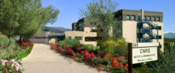
- Institut de Pharmacologie Moléculaire et Cellulaire (IPMC) - Sophia Antipolis
-
IPMC UMR7275/UNS
660 Route des Lucioles
Sophia Antipolis
06560 ValbonnePERSONNEL
Samah Rekima
Ingénieur service Imagerie
rekima@ipmc.cnrs.fr
04.93.95.77.83-87-88-89Sophie Abélanet
Ingénieur service Imagerie
abelanet@ipmc.cnrs.fr
04.93.95.77.83-87-88-89EQUIPEMENTS
Microscopie plein champ :
- Axioplan2 Imaging Carl Zeiss (2003) - Caméra Hamamatsu N&B CCD Orca ER - Logiciel µManager - Droit
- Axiovert200M Carl Zeiss (2003) - Caméra Hamamatsu sCMOS Orca Flash 4.0 - platine motorisée - Logiciel Metamorph - Inversé
- Axio Observer Carl Zeiss (2007) - Caméra EMCCD Cascade 512 Photometrics - Logiciel MetaFluor - Inversé
Microscopie plein champ automatisée :
- Cytation 5 Biotek (2015) - Caméra Grasshopper CCD - Logiciel Gen5 - Lecteur de plaque - Inversé
- Image Xpress pro Molecular Devices (2023)
Micro-dissecteur laser :
- Microdissecteur PALM Carl Zeiss sur AxioObserver Z1 - Caméra couleur MRC5 - Caméra N&B mRm - platine motorisée (piloté par Axiovision et Palmrobo) - Inversé
Microscopie confocale :
- LSM780 Carl Zeiss (2012) - détecteur spectral et GaAsp - Logiciel ZEN - Inversé
- TCS SP5 Multi-Photon Leica Microsystems (2012/2020) - détecteurs hybrides NDD- surplatine piezo - scanner résonant - Laser Chameleon UltraII et OPO- Droit
- TCS SP8 STED Leica Microsystems (2017) - détecteur hybride - surplatine piezo - scanner résonant - Inversé
Spinning disk :
- Spinning Disk UltraviewVox Perkin Elmer (2010) - Eclispse TiE Nikon - Caméra Hamamatsu EM-CCD - Logiciel Volocity - Inversé
Feuille de lumière :
- Ultramacroscope à feuille de lumière Home-Made sur MVX10 Olympus (2014) - Caméra sCMOS Flash4 Hamamatsu - Banc 4 lasers Oxxius - Logiciel Inscoper
Logiciels d'analyses :
- Volocity 6.3 (PerkinElmer)
- Imaris 9.6 pour analyses 3D (Bitplane)
- Huygens Core/Huygens Remote Manager pour la déconvolution (SVI)
- Institut de Biologie de Valrose (iBV) - Nice
-
UMR CNRS 7277- Inserm U1091- UNS
Faculté des sciences - Parc Valrose
06108 Nice cedex 2PERSONNEL
Sameh Ben Aïcha
Responsable service Imagerie (PRISM)
sameh.ben-aicha@univ-cotedazur.fr
04.89.15.06.80Robert Arkowitz
Responsable scientifique
Robert.ARKOWITZ@univ-cotedazur.fr
04 89 15 0740Maximilian Furthauer
Responsable scientifique
maximilian.furthauer@univ-cotedazur.fr
04.89.15.08.35EQUIPEMENTS
Microscopie plein champ :
- Axiocolor : AxioPlan Carl Zeiss / Caméra couleur pour coloration
- Axiofluo : AxioPlan Carl Zeiss / Caméra noir & blanc pour la fluorescence
Vidéo-microscopie :
- Videobserver : AxioObserver Carl Zeiss (2011) - Caméras Andor EMCCD iXion 897 et sCMOS Neo - Logiciel Metamorph
- Video-microscope Axiovert200M Carl Zeiss (2004) - Caméra Andor sCMOS Neo - Logiciel Metamorph
- Lattice SIM Elyra7 Lattice SIM Carl Zeiss (2023)
Microscopie confocale :
- TCS SPE Leica Microsystems (2007)
- TCS SP5 Leica Microsystems (2011) - détecteur hybride - surplatine piezo - scanner résonant - Inversé
- LSM710 Carl Zeiss (2012) - détecteur spectral - Inversé
- LSM780 Carl Zeiss (2012) - détecteur spectral et GaAsp - laser Bi-photon (MaiTai) - détecteur NDD en fluorescence et transmission
- LSM880 Carl Zeiss (2018) - Fast Airyscan - détecteur interne GaAsp - Surplatine piezo - Inversé
- LSM980 Carl Zeiss (2024)
Spinning disk :
- Spinning disk: Olympus/Andor/Yokogawa X1 (2010) - Module Dual cam (EM-CCD) - 3D rapide - surplatine piezo
- SPIN-FRAP-TIRF: Nikon TiE/Yokogawa W1 (2016) - module de photomanipulation (Ilas2) - caméra sCMOS zyla 4.2+
Feuille de lumière :
- Lightsheet Z7 Carl Zeiss (2023)
Logiciels d'analyses :
- Imaris 9.6 pour analyses 3D (Bitplane)
- Huygens Core/Huygens Remote Manager pour la déconvolution (SVI)
- Centre Méditerranéen de Médecine Moléculaire (C3M) - Nice
-
C3M
Bâtiment Universitaire ARCHIMED
151 route Saint Antoine de Ginestière
BP 2 3194
06204 Nice Cedex 3PERSONNEL
Marie Irondelle
Responsable service Imagerie
marie.irondelle@univ-cotedazur.fr
04.89.06.42.58Anne Doye
Ingénieur service Imagerie
anne.doye@univ-cotedazur.fr
04.89.06.42.63EQUIPEMENTS
Microscopie plein champ :
- Eclipse Ci-S Nikon (2016) - pour coupe histologique - droit
Vidéo-microscopie :
- Axiovert 200M Carl Zeiss (2006) - Caméra Coolsnap HQ2 Photometrics - platine motorisée - Logiciel Metamorph - Inversé
- AM TIRF Leica Microsystems (2007) - Caméra DFC360FX - platine motorisée - Logiciel LAS AF - Inversé
Micro-dissecteur laser :
- Microdissecteur PALM Zeiss (2010) - Caméra couleur MRC5 - Caméra N&B mRm - platine motorisée (piloté par Axiovision et Palmrobo) - inversé
Microscopie confocale :
- Nikon A1R (2015) : scanner résonant, module d'automatisation des acquisitions, maintien tde focus, mosaïques
Logiciels d'analyses :
- Imaris 9.6 pour analyses 3D (Bitplane)
- Huygens Core/Huygens Remote Manager pour la déconvolution (SVI)
- Institut de la Mer de Villefranche (IMEV) - Villefranche
-
Observatoire Océanologique de Villefranche-sur-Mer
Port de la Darse
181, Chemin du Lazaret
06230 Villefranche sur mer CedexPERSONNEL PIM - service Imagerie
Sébastien Schaub
Responsable service Imagerie (PIM)
sebastien.schaub@imev-mer.frEQUIPEMENTS
Microscopie plein champ manuelle :
- BX51 Olympus (2013) - Camera Axiocam 506 color Carl Zeiss (brightfield, Darkfield, fluo) - Droit
- Imager A2 Zeiss (2014) - Camera Axiocam 506 color Carl Zeiss (brightfield, Darkfield, fluo) - Droit
- Stéréomicroscope Zeiss Stemi 508 (2019) - Caméra AxioCam 305 color Carl Zeiss (brightfield, oblique)
- Stéréomicroscope M205FA Leica Microsystems (2014) - Caméra MC170 HD Leica Microsystems (brightfield,oblique, fluorescence)
Microscopie plein champ motorisée :
- Axiovert200M Carl Zeiss (2013) - Camera Fusion BT Hamamatsu (brightfield, fluo) - Logiciel Metamoprh - Inversé
- AxioObserver 7 Carl Zeiss (2019) - Caméra Flash 4 v3 Hamamatsu - Logiciel ZEN Blue - Inversé
Microscopie confocale :
- TCS SP8 Leica Microsystems (2015) - Détecteurs hybrides - Inversé
- Stellaris 5 Leica Microsystems (2020) - Laser blanc - Tau Sense
Feuille de lumière :
- Lightsheet LS2 Viventis / Leica Microsystems (2023)
Microscopie électronique à balayage :
- Microscope électronique environnemental Hitashi 4000+ (2021)
Logiciels d'analyses :
- Imaris 10.0 pour analyses 3D (Bitplane)
- Huygens Core/Huygens Remote Manager pour la déconvolution (SVI)
- MATLAB
PERSONNEL PIQV - service d'imagerie quantitative
Manoela Brandão
Responsable service Imagerie quantitative (PIQV)
manoela.brandao@imev-mer.fr
04.93.76.38.51Marc Picheral
Responsable scientifique (PIQV)
marc.picheral@imev-mer.fr
04.93.76.38.08EQUIPEMENTS PIQV - service d'imagerie quantitative
Acquisition de plancton :
- 2 Imaging FlowCytobot (IFCB)
- 4 FlowCam
- 7 ZooScan
- 4 Underwater Vision Profiler (UVP5)
- 18 Underwater Vision Profiler 6 Low Power (UVP6-LP)
- 4 Underwater Vision Profiler 6 High Frequency (UVP6-HF)
Logiciels d'analyses:
Zooprocess
- Centre Commun de Microscopie Appliquée (CCMA) - Nice
-
CCMA
UFR Sciences
Parc Valrose
28, avenue Valrose
06108 Nice Cedex 2PERSONNEL
Sandra Lacas-Gervais
Responsable scientifique service imagerie électronique
sandra.lacas-gervais@univ-cotedazur.fr
04.89.15.00.92François Orange
Ingénieur responsable technique du MEB
francois.orange@univ-cotedazur.fr
04.89.15.00.93Sophie Pagnotta
Ingénieur responsable technique du MET
sophie.pagnotta@univ-cotedazur.fr
04.89.15.00.90Christelle Boscagli
technicienne responsable équipements et préparation des échantillons
christelle.boscagli@univ-cotedazur.fr
04.89.15.00.94EQUIPEMENTS
Microscopie plein champ :
- Thunder Imager 3D sur DMI8 Leica Microsystems (2020) - computational clearing et déconvolution intégrée - Inversé
- Institut de Recherche sur le Cancer et le Vieillissement (IRCAN) - Nice
-
UMR CNRS 7284- INSERM U1081-UNS
Faculté de Médecine Pasteur
Avenue de Valombrose
06107 NICE Cedex 2PERSONNEL PICMI
Soline Estrach
Responsable scientifique service imagerie (PICMI)
soline.estrach@univ-cotedazur.fr
04.89.15.36.91Aaron Mendez-Bermudez
Ingénieur service imagerie (PICMI)
aaron.mendez-bermudez@univ-cotedazur.frFrédéric Brau
Responsable des plateformes et Ingénieur service imagerie (PICMI)
frederic.brau@cnrs.frEQUIPEMENTS PICMI
Microscopie plein champ :
- AxioImager Z2 Zeiss (2011) - Caméra sCMOS AxioCam705 mono - Platine motorisée 8 lames - Logiciel ZEN - Droit
- DM4000B Leica Microsystems - Caméra couleur DFC 425C - Droit
Vidéo-microscopie :
- AxioObserver Z1 Carl Zeiss (2010) - Caméra couleur Zeiss AxioCam Icc1 et Hamamatsu sCMOS Orca Flash 4.0 LT C11440 - Platine motorisée - Contrôle CO2 et température - Logiciel ZEN - Inversé
- Axiovert 200M Carl Zeiss - Caméra Photometrics EMCCD Cascade II - Platine motorisée - Contrôle CO2 et température - Logiciel Metamorph - Inversé
- DeltaVision Elite TIRFM Olympus (2013) - Caméra sCMOS PCO Edge 5.5- Platine motorisée - Contrôle CO2 et température - Logiciel SoftWorx - Inversé
Microscopie confocale :
- LSM800 Carl Zeiss (2018) - Platine motorisée - Logiciel ZEN - Droit
- LSM880 Zeiss (2018) - Fast Airyscan - détecteur spectral GaAsp - Platine motorisée - Surplatine piezo - Logiciel ZEN - Inversé
Feuille de lumière :
- Lightsheet Z1 Carl Zeiss (2018)
Logiciels d'analyses :
- Imaris 9.6 pour analyses 3D (Bitplane)
- Huygens Core/Huygens Remote Manager pour la déconvolution (SVI)
- Institut Sophia Agrobiotech INRAE (ISA) - Sophie Antipolis
-
Institut Sophia Agrobiotech INRAE PACA
400 route des chappes
BP 167
06903 Sophia Antipolis CedexPERSONNEL
Olivier Pierre
Responsable service imagerie (SPIBOC)
olivier.pierre@inrae.fr
04.92.38.64.58Julie Soltys
Ingénieur service imagerie (SPIBOC)
julie.soltys@inrae.fr
04.92.38.64.58Janice de Almeida-Engler
Responsable scientifique service imagerie (SPIBOC)
janice.de-almeida@inrae.fr
04.92.38.64.59EQUIPEMENTS
Microtomes :
- Vibratome VT1200S Leica
- Microtome HM360-Microm Microtech
- Microtome Rotatif-Jung
Loupes binoculaires :
- MZFLII Leica Microsystems- Caméra AxiocamICc1 - Logiciel ZEN
- StéréoDiscvoveryv12 Carl Zeiss – Caméra couleur CCD AxioCam - Logiciel ZEN
Microscopie plein champ :
- VHX-2000 Keyence (2013) - digital microscope - CCD color camera with 20x to 6000x magnification capacity - Logiciel Communication Software
- AxioZoom Carl Zeiss (2018) - module Apotome2 - motorized stage with Orca Flash4LT & color coded sCMOS AxioCam305
- AxioImagerZ1 Carl Zeiss (2007) - Apotome2- Platine motorisée - Caméra N&B Orca Flash4LT Hamamatsu - Caméra N&B AxioCam MRm - Logiciel ZEN - Inversé
- AxioPlan Carl Zeiss (2007) - Caméra couleur sCMOS Axiocam705 - Logiciel ZEN - Droit
Microscopie confocale :
- LSM880 Carl Zeiss (2015 upgrade 2017) - Fast Airyscan - Détecteurs GaAsp - Inversé
Logiciels d'analyses :
- Amira Extension XImagePAQ (Thermo Fisher)
- Huygens Core/Huygens Remote Manager pour la déconvolution (SVI)